



Microscopic root canal treatment is a form of endodontic therapy performed with the aid of a dental operating microscope, which magnifies the root canal system up to 25 times its actual size. This level of magnification allows the dentist to locate hidden canals, detect micro-cracks, and clean the root system more thoroughly — all of which reduces the risk of failure and re-treatment.
If you’ve been told you need a root canal and are weighing your options, understanding what microscope endodontics actually offers can help you make a more informed decision.
What Is Microscopic Root Canal Treatment?
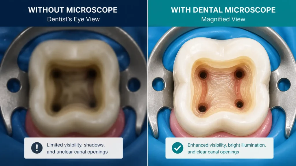
Microscopic root canal treatment — also called microscope-assisted RCT or microscope endodontics — is root canal therapy performed under the magnification of a surgical operating microscope. Unlike traditional RCT, which relies on tactile feel and X-rays, microscopic RCT gives the treating dentist a clear, illuminated, magnified view of the entire working field inside the tooth.
Root canals are narrow, curved, and often branching. Many treatment failures in conventional RCT occur because a canal was missed, a perforation went undetected, or calcified tissue blocked access. The dental microscope addresses each of these problems directly.
How a Dental Microscope Works During RCT
The dental operating microscope is ceiling-mounted or stand-mounted beside the dental chair. It uses coaxial illumination — meaning the light source is aligned with the line of sight — so there are no shadows inside the tooth’s interior.
The dentist views the tooth through the eyepiece at magnifications ranging from 4× to 25×. At lower magnification, the overall tooth structure is visible. At higher magnification, individual canal orifices, dentinal cracks, and even the junction between the pulp floor and the canals come into clear view. Most procedures also record footage, which can be shown to patients and referred to during treatment planning.
Microscopic vs Conventional Root Canal — What Actually Changes?
Both procedures aim to remove infected pulp tissue, shape the canal, and seal the tooth. The difference lies in the quality of vision available at every step.
| Feature | Conventional RCT | Microscopic RCT |
| Magnification | Naked eye / dental loupe (2–4×) | Operating microscope (4–25×) |
| Canal detection | Standard X-ray guided | Visual + X-ray; missed canals rarely occur |
| Crack detection | Often missed | Fine cracks and fractures visible |
| Tissue removal | Estimated | Conservative and precise |
| Risk of perforation | Moderate | Significantly lower |
| Suitability for re-treatment | Limited | High — ideal for re-treatment cases |
| Documentation | X-rays | X-rays + intra-procedure video |
| Operator fatigue | Higher in complex cases | Lower — better posture and vision |
The difference is most significant in complex cases: calcified (blocked) canals, curved roots, re-treatment of failed conventional RCT, and teeth with unusual anatomy.
Who Needs Microscopic Root Canal Treatment?
Not every root canal requires a microscope, but certain clinical situations benefit enormously from one.
Microscope endodontics is especially recommended when:
- A previous root canal has failed and re-treatment is being planned
- The canals are calcified or narrowed due to age or previous trauma
- The tooth has complex anatomy (extra canals, curved roots)
- A fractured instrument was left inside the canal during a prior procedure
- There is suspected root perforation that needs to be located and repaired
- The treating dentist needs to assess whether a vertical root fracture is present
- Standard techniques have not produced the expected outcome
For most straightforward first-time RCTs in single-canal teeth with normal anatomy, experienced operators can achieve good results with conventional techniques. However, even in routine cases, precision root canal under microscopy provides a higher standard of care.
What Happens During the Procedure?
Microscope-assisted root canal treatment follows the same fundamental steps as conventional RCT. The difference is the level of detail at which each step is performed.
- Local anaesthesia — The area around the tooth is numbed. This is the same as conventional RCT. Patients typically feel pressure but no pain.
- Rubber dam placement — A thin rubber sheet isolates the tooth, keeping the field clean and the patient safe from irrigant solutions.
- Access opening — The dentist creates an opening through the crown of the tooth to reach the pulp chamber. Under the microscope, this opening is made with precision, preserving as much healthy tooth structure as possible.
- Canal identification — All canal orifices are located. Under magnification, canals that are invisible to the naked eye become clearly visible — a critical advantage in teeth with extra canals (such as upper first molars, which frequently have four canals instead of the expected three).
- Canal shaping and cleaning — Rotary files are used to shape the canal to a taper that allows thorough irrigation. Irrigants (usually sodium hypochlorite and EDTA) flush out bacteria and tissue debris.
- Final inspection — The clean canal walls are inspected under high magnification for remaining tissue, micro-cracks, or accessory canals.
- Obturation — The canals are filled with gutta-percha and a sealer to prevent reinfection.
- Restoration — A temporary or permanent restoration seals the access opening. In most cases, a crown is recommended afterward to protect the treated tooth.
Total chair time is typically 60–90 minutes per session, depending on the number of canals and the complexity of the case.
Does Microscopic Root Canal Hurt More Than Regular RCT?
No. The procedure is performed under local anaesthesia, and the experience from the patient’s perspective is essentially the same as conventional RCT. Most patients report feeling pressure and vibration during the procedure but no sharp pain.
In fact, many patients find that microscope-assisted procedures are faster and more comfortable because the dentist works with greater efficiency — spending less time feeling for canals and more time confirming that each step is complete. There is no additional injection, no different anaesthesia protocol, and no increased sensitivity associated with the microscope itself.
Recovery: What to Expect After Microscope-Assisted RCT
Recovery after microscopic root canal treatment mirrors recovery after standard RCT. The treated tooth and surrounding gum may feel tender for one to three days. This is normal — the periapical tissues (around the root tip) respond to the cleaning process with mild inflammation.
What to expect in the first 72 hours:
- Mild to moderate soreness when biting or pressing on the tooth
- Occasional aching sensation, more pronounced on day one
- Slight gum sensitivity around the treated area
Managing discomfort:
- Over-the-counter analgesics such as paracetamol or ibuprofen are usually sufficient
- Avoid chewing hard foods on the treated side until the permanent crown is placed
- Maintain gentle oral hygiene — continue brushing and flossing normally
Most patients return to work and normal activity the same day or the following day. If pain intensifies after 72 hours rather than subsiding, contact your dentist, as this can occasionally indicate a need for further evaluation.
Myths vs Facts About Microscopic Root Canal
| Myth | Fact |
| “Microscopic RCT is only for complicated cases.” | While it excels in complex cases, the precision it offers benefits any root canal procedure. |
| “It’s experimental technology.” | Dental operating microscopes have been in clinical use for over three decades and are endorsed by the American Association of Endodontists. |
| “The microscope makes the procedure longer.” | Initial set-up adds a few minutes, but working under magnification often reduces overall procedure time by improving efficiency. |
| “The results are the same as regular RCT.” | Studies consistently show lower rates of missed canals, perforations, and treatment failures with microscope-assisted endodontics. |
| “It’s too expensive to be worth it.” | Given that re-treatment or tooth extraction and implant placement cost significantly more, the upfront investment in precision often saves money long-term. |
| “All dentists offer this.” | Microscope endodontics requires specific training and equipment. Not all clinics in South Kolkata currently offer it. |
Frequently Asked Questions
Q1: What is microscopic root canal treatment? Microscopic root canal treatment is endodontic therapy performed using a high-powered dental operating microscope. The microscope magnifies the root canal system up to 25 times, allowing the dentist to see hidden canals, detect micro-cracks, and clean the root system with a level of precision not possible with the naked eye or standard dental loupes.
Q2: Is microscopic root canal more painful than regular RCT? No. Both procedures use local anaesthesia. Patients feel pressure but not pain during the procedure. Post-procedure discomfort is also comparable — mild soreness for one to three days, managed easily with standard analgesics.
Q3: How long does microscopic root canal take? A single-session microscopic root canal typically takes 60 to 90 minutes. Complex cases, re-treatments, or teeth with many canals may require two sessions. Your dentist will give you a more specific estimate after examining your tooth and X-rays.
Q4: Who is a good candidate for microscopic RCT? Anyone requiring root canal treatment can benefit. It is especially recommended for patients undergoing re-treatment, those with calcified or curved canals, teeth with unusual anatomy, and cases where a conventional RCT has not resolved the infection.
Q5: What is the success rate of microscopic root canal? Success rates vary by case complexity, but microscope-assisted RCT consistently outperforms conventional techniques, particularly in re-treatment and complex cases. Studies report success rates of 85–95% for primary treatments under microscopy.
Q6: Will I need a crown after microscopic root canal? In most cases, yes. A crown protects the tooth from fracture after the pulp — which supplies moisture to the tooth — has been removed. Your dentist will advise you based on how much natural tooth structure remains after treatment.
Q7: Is microscopic root canal available in South Kolkata? Yes. City Smiles Dental Care in South Kolkata offers microscopic root canal treatment. Patients from Garia, Baghajatin, Jadavpur, Tollygunge, Santoshpur, and Dhakuria regularly visit the clinic for advanced endodontic care. You can book an appointment here.
Q8: How does microscope endodontics reduce re-treatment rates? Missed canals are the leading cause of root canal failure. Under microscope magnification, even accessory canals and lateral canals invisible to the naked eye can be identified and treated. This thoroughness dramatically reduces the chance of residual infection driving re-treatment.
Q9: Can microscopic RCT save a tooth that a previous root canal failed on? Yes. Microscope-assisted re-treatment is one of the primary applications of dental microscopy. The microscope helps locate missed canals, remove old filling material, repair perforations, and retrieve broken instruments — all of which are common challenges in re-treatment cases.
Q10: How should I prepare for a microscopic root canal appointment? No special preparation is needed. Eat a normal meal before your appointment, as local anaesthesia will make chewing uncomfortable for a few hours afterward. Arrange transport if you feel anxious about dental procedures, as some patients prefer mild sedation. Bring any prior dental X-rays or treatment records, especially if this is a re-treatment case.
Getting Microscopic Root Canal Treatment in South Kolkata
For patients across South Kolkata — whether you’re in Garia, Baghajatin, Jadavpur, Tollygunge, Santoshpur, or Dhakuria — finding a clinic equipped for microscope endodontics can feel uncertain. Advanced RCT technology is not universally available at every practice, and the skill of the operator matters as much as the equipment itself.
At City Smiles Dental Care, microscopic root canal treatment is performed using a clinical-grade dental operating microscope by trained dental professionals with experience in precision endodontics. Every case begins with a thorough clinical and radiographic assessment so that treatment is planned accurately before the first appointment.
If you’ve been told you need a root canal — or if a previous root canal has not resolved your symptoms — a microscopic evaluation can provide answers that conventional imaging sometimes cannot.
Conclusion
Microscopic root canal treatment represents a meaningful upgrade in how endodontic care is delivered. The dental microscope doesn’t change what root canal therapy is — it changes how well it can be done. Hidden canals are found. Cracks are caught early. Healthy tissue is preserved. And the chance of needing re-treatment is significantly reduced.
For patients in South Kolkata who want precision root canal treatment without travelling far, City Smiles Dental Care offers microscope-assisted endodontic care with the attention to detail that complex cases demand and routine cases deserve.
Book Your Microscopic Root Canal Consultation
If you’re experiencing tooth pain, have been advised to undergo root canal treatment, or are looking for a second opinion on a failed RCT, we’re here to help.
Book an Appointment at City Smiles Dental Care →
You can also find us on Google Maps and read patient reviews on our Google Business Profile to learn more about the care experience at our clinic.
Serving patients from Garia, Baghajatin, Jadavpur, Tollygunge, Santoshpur, Dhakuria, and all of South Kolkata.